


![]() n the 1980s, Continental Europe started to contribute intensively to MR imaging. Rapid imaging originated in European laboratories.
n the 1980s, Continental Europe started to contribute intensively to MR imaging. Rapid imaging originated in European laboratories.
Jürgen Hennig (Figure 20-46), together with A. Nauerth and Hartmut Friedburg, from the University of Freiburg in Germany introduced RARE (Rapid Acquisition with Relaxation Enhancement) imaging in 1986 [⇒ Hennig 1986]. This technique is probably better known under the commercial names of 'fast' or 'turbo' spin-echo (Figure 20-47).
 Figure 20-46: Jürgen Hennig
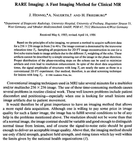
Figure 20-47:
First page of Hennig's article about RARE imaging (submitted in 1985, published in 1986).
The beginning of the article summarizes the problem to be solved:
"Conventional imaging techniques used in MRI take several minutes for a multiple and/or multiecho 256×256 image. The use of these time-consuming methods causes several problems in routine clinical work. These well known problems include patient discomfort and positioning …"
At about the same time, FLASH (fast low angle shot) appeared, opening the way to similar gradient-echo sequences. FLASH had a completely different approach and, for non-scientific reasons, was very rapidly adopted commercially. The FLASH sequence was developed at Max-Planck-Institute, Göttingen, by Axel Haase (Figure 20-48), Jens Frahm (Figure 20-49), Dieter Matthaei, Wolfgang Hänicke, and Dietmar K. Merboldt [⇒ Haase 1986].
 Figure 20-48: Axel Haase
 Figure 20-49: Jens Frahm
The inclusion of Hennig's RARE into the clinical imaging protocols was slower, and Mansfield's echo-planar imaging (EPI) took even more time to find its way into clinical imaging — for technical reasons.
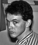 Figure 20-50: Richard A. Jones
Acquiring images faster and with better quality remained one of the main goals in MR research. New ideas and distinct concepts were developed, for instance k-space substitution as proposed by Richard A. Jones (Figure 20-50) [⇒ Jones 1993].
A combination of dedicated hardware and specific software led to parallel imaging which can reduce imaging time considerably. A first technique was described by Sodickson and Manning but it required a particular coil configuration [⇒ Sodickson 1997].
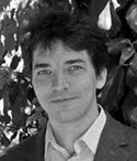 Figure 20-51: Klaas Prüssmann
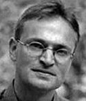 Figure 20-52: Markus Weiger
In 1999 Klaas Prüssmann (Figure 20-51) and Markus Weiger (Figure 20-52) introduced SENSE and thus offered a more general solution [⇒ Pruessmann 1999]. Algorithms of the GRAPPA type, introduced a year later by Mark A. Griswold [⇒ Griswold 2002], work better than the SENSE type for abdominal and thoracic or for echo planar imaging.


|

|
|---|