


 efinition. Molecular imaging is a catchword and misnomer used as an umbrella term for all biological and medical imaging technologies and methods that aim at picturing anatomy, histology as well as features and processes at the cellular and molecular level [⇒ James 2012, ⇒ Weissleder 2015]. It is basically an extension of radioisotope tracer scanning and imaging into other fields of medical imaging.
Commonly, molecular imaging is just a synonym of contrast-enhanced imaging. Molecular imaging media are contrast agents of one kind or another (plain contrast enhancers, static tracers, or responsive agents).
Given the increasing understanding of molecular mechanisms of disease and the development of innovative therapies at the genetic level, molecular imaging is aimed at the exploitation of specific molecules as the source of image contrast (Figure 13-20). However, although MR imaging does not possess sufficient sensitivity to image single molecules, responsive MR agents can dynamically change one or more of their physico-chemical properties when interacting with their intended molecular biomarker.
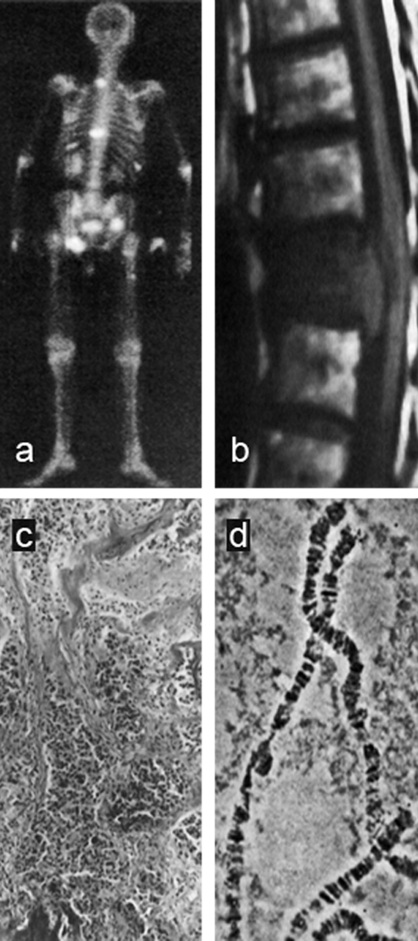
Figure 13-20:
(a) Whole body screening: radioisotope scan;
(b) organ level: MR spine study without contrast enhancement;
(c) tissue level: bone histology;
(d) genetic level: chromosome screening.
For the time being, clinical molecular imaging will stay in the realm of radioisotope scanning and contrast-agent enhanced imaging, sometimes related to hybrid systems such as PET-MRI or PET-CT. A good review of responsive MR agents was published by Hingorani et coll. [⇒ Hingorani 2015].
At the organ level, diagnostic imaging today is able to visualize gross parameters of disease and to describe, e.g., tumor burden. In the future, it will be possible to mark tumors and to target drugs as well as contrast agents. At the genetic level researchers hope to highlight genetic mutations and thus perform gene targeting and therapy.
Researchers will try to leave some of the existing pathways: instead of looking for relatively gross parameters of disease, they will try to explore beyond the tissue level on the cell or even the molecular level. They will try to connect diagnosis with therapy; one of the main goals being the imaging assessment of therapeutic effectiveness at the molecular level, long before phenotypic changes occur [⇒ Weissleder 2015].
MRI and Molecular Imaging. Whereas the spatial resolution of MR imaging is very good, its sensitivity is low and thus limiting. Present software and hardware improvements, such as more sophisticated coil design, seem not to be able to bridge the existing gap.
Any increase of field strength beyond today's clinical ultrahigh fields (3 or even 7 Tesla) to gain crucial signal strength is restricted by the specific absorption rate (SAR) and by side effects of the magnetic fields.
A detour would possibly be the use of hyperpolarization; however, hyperpolarized clinical ventilation imaging was not promising and mostly abandonded for commercial reasons.
MEMRI (manganese-enhanced MRI) of the heart is a good example of one of the few promising molecular imaging methods, because it may be possible to use the same manganese-based compound for diagnostics and treatment of, e.g., myocardial infarctions, cancer, and drug intoxication (theragnostic properties). It is inexpensive and addresses a mass market.
Intelligent and/or responsive agents might be feasible to specific applications in magnetic resonance imaging. They could alter their relaxivity according to local temperature, pH, or chemical activity.
Yet, there are a lot of uncertainties in this kind of research, and with the advent of in vitro screening techniques such as easy-to-use blood tests for Alzheimer's disease, molecular imaging will be increasingly challenged.
It will, however, remain one of the major R&D topics in diagnostic and therapeutic imaging and contrast agent research of the coming decades.
Molecular imaging — some more thoughts …


|

|
|---|
