


 he magnetic field produced by an electron is much stronger than that produced by a proton. However, in most substances the electrons are paired, resulting in a weak net magnetic field. As seen in Table 13-01, gadolinium with its seven unpaired electrons is best suited for this purpose, followed by manganese.
Paramagnetic contrast agents, with the exception of dysprosium-based preparations, are called positive agents. Their effect on T1 and T2 is similar, but since T1 of tissues is much higher than T2, the predominant effect at low doses is that of T1 shortening. The principle was shown in Figure 04-05.
Thus, tissues taking up such agents will become bright when using a T1-weighted sequence (Figure 13-02 and Figure 13-03).
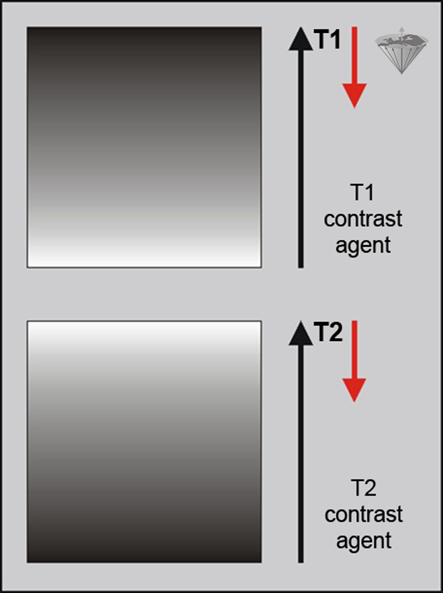
Figure 13-02:
Influence of positive (T1) and negative (T2, T2*) MR contrast agents upon signal intensity. Paramagnetic agents are mainly used to shorten T1 relaxation and thus brighten the region of interest, whereas ferro- and superparamagnetic agents shorten T2 and T2* and thus darken the image (red arrows).
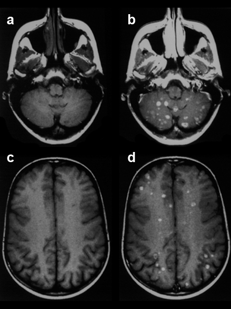
Figure 13-03:
This figure gives an example of a clinical case where finally the application of such a positive contrast agent helped the diagnostic process and showed the extent of the disease: Patient with breast cancer and recent neurological symptoms. T1-weighted images. The plain MR images (precontrast: a and c) do not reliably reveal brain lesions. However, the contrast-enhanced MR images (postcontrast: b and d) show a large number of metastases.
Negative contrast agents influence signal intensity usually by shortening T2* and T2. This darkens the region of interest (Figure 13-02 and Figure 13-04).
If one reduces their size, they lose their permanent magnetic characteristics and are then called superparamagnetic particles [⇒ Weissleder 1992]. Depending on their particle size and coating, these compounds can also become T1 agents.
Magnetite, Fe3O4, is such a superparamagnetic particle. Coated with an inert resin, magnetites can be used for oral or intravenous applications (cf. iron oxides).
Ferro- and superparamagnetic contrast agents produce local magnetic field gradients which disrupt the homogeneity of the local magnetic field. T2 is reduced due to the diffusion of water through these field gradients. However, their principal effect is a reduction in T2*. For this reason, the effects of such contrast agents are best observed using GRE sequences where T2* effects are retained. This kind of effect is referred to as susceptibility effect. It is dependent on magnetic field strength, with the effect increasing as the square of the magnetic field strength.
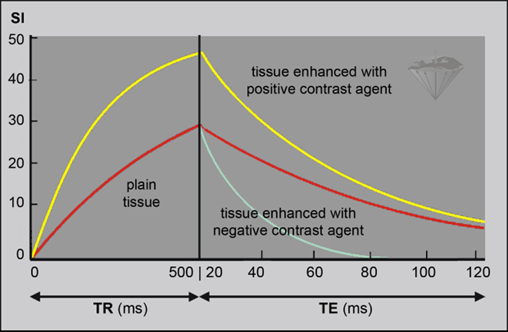
Figure 13-04:
Influence of a positive (T1) and negative (T2, T2*) contrast agent upon signal intensity (SI).
T1 relaxation is accelerated by positive agents, and the spins recover faster. Therefore, at a given TR, signal intensity (yellow curve) is higher than in the same tissue without contrast agent (red curve). Only a T1-weighted sequence (in this case a spin echo sequence) with short TE highlights this contrast enhancement.
With negative contrast agents, T2 relaxation is accelerated; signal intensity is lower (light blue curve; in the "TR" part of the graph this curve overlaps with the red curve).


|

|
|---|