


 ultispectral images of the same body region can simply be overlaid to give an impression of the exact location of certain contrast-enhanced structures (data fusion). Usually, a high spatial resolution T1-weighted MR image is used as a background image to show the anatomical structures and the contrast-enhanced image is projected onto this picture.
In MR imaging this has been first shown with perfluorinated ventilation images in 1982 (Figure 15-07) [⇒ Rinck 1983].
Today the method is used as a multiparametric or multimodal fusion of data as well as longitudinal (time domain) integration of single modality data, commonly applied in fMRI studies or in intermodality comparison between, e.g., CT or MR and PET images. Here, the information obtained from PET is overlaid or imprinted onto the more detailed anatomic images acquired with MR imaging or CT.
 Practical Applications. The method is useful since it allows a better visualization to locate certain processes. The implementation is relatively simple.
It is mainly applied as an auxiliary tool to facilitate visualization of enhancement visible on postcontrast images (Figure 15-08), but also in MR angiography to highlight veins after subtraction of the CE-MRA images of the arterial phase, and in MRSI (Figure 05-08).
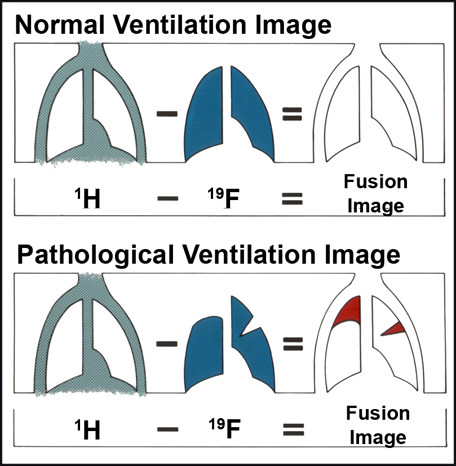
Figure 15-07:
Diagram explaining the theory of the first fusion images in MRI (1982). Subtraction of hydrogen and fluorine images of the lungs (cf. Figure 20-40).
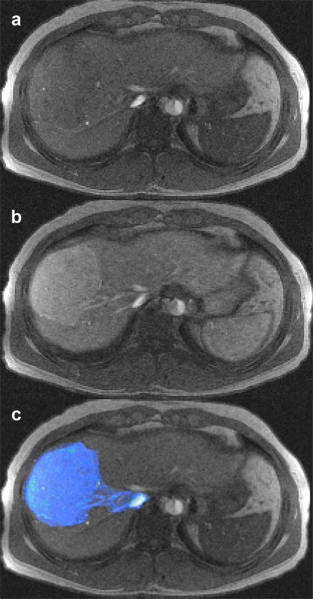
Figure 15-08:
Multichannel images: (a) plain image of a liver, (b) contrast-enhanced liver, with depiction of focal nodular hyperplasia, part of a dynamic series, and (c) overlay (superposition) of enhanced lesion on plain liver image.


|

|
|---|