


 completely different approach to rapid imaging was used by the first pulse sequences, which shortened imaging time in routine clinical settings. The generic name of these sequences is gradient-echo (GE) sequences or, better, gradient-recalled echo (GRE) sequences, and they come in a plethora of different acronyms. The basics of GRE were explained in Chapter 6.
The first sequence in this group was presented in 1986 by Axel Haase and collaborators and dubbed FLASH [⇒ Haase 1986]. The FLASH (Fast Low Angle Shot) sequence is a saturation recovery sequence with a short repetition time (TR ≤200 ms), a low flip angle (≤90°), and a gradient echo for refocusing.
The application of flip angles different from 90° and 180° brought an end to the ideology of long waiting times, which was based upon the belief that T1 is the limiting time factor of MR imaging. The reason for using a low flip angle is illustrated in Figure 08-05.
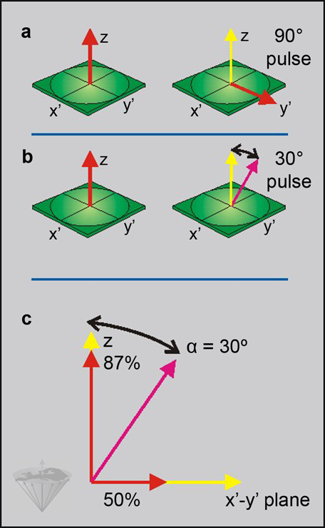
Figure 08-05:
Principles of (a) a standard pulse sequence, compared to (b) a rapid imaging sequence of the FLASH type. In both cases, the net magnetization during equilibrium is aligned with the z-axis. In the standard sequence, a 90° pulse tilts the magnetization into the x'-y' plane. No longitudinal component remains. In the FLASH-type sequence, a flip angle α ≤90° is applied. Such a pulse divides the magnetization in transverse and longitudinal components.
(c) In our example α equals 30°. This results in a reduction of the longitudinal magnetization to 87%, whereas the transverse magnetization is 50% of the available longitudinal magnetization.
The flip angle which will give maximum signal is known as the Ernst angle.
When a 90° flip angle is applied, we convert all of the longitudinal magnetization (in the z-axis) into transverse magnetization (signal in the x'-y' plane), while, e.g., for a 30° flip angle the amount of transverse magnetization is halved (sin 30°), but we still have 87% of the z-magnetization (cos 30°). The z-magnetization will recover at a rate determined by T1 during the interpulse interval. However, since the TR is short in FLASH sequences, the z-magnetization left by the previous pulse becomes dominant and significantly increases the signal obtained after the next RF pulse.
For a given repetition time, the flip angle which will give maximum signal can be calculated. It is known as the Ernst angle [⇒ Ernst 1966]:
where TR is the repetition time and T1 the longitudinal relaxation time.
Figure 08-06 summarizes the main differences between a spin-echo and a gradient-echo (FLASH) pulse sequence.
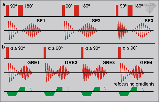
Figure 08-06:
Principles of (a) a standard pulse sequence, compared to (b) a rapid imaging sequence of the FLASH type.
(a) In the spin-echo pulse sequence, the echo is created by a 180° pulse. This involves relatively long time delays and high power deposition in the examined sample. Because of the dependence of TR on T1, TR has to be relatively long. (b) In the FLASH sequence, any pulse angle can be used instead of the initial 90° pulse. The echo is formed by gradient switching. This can be done faster and with less power deposition (potentially less hazardous for the patient). Thus, TR (and TE) can be shortened.
SE = spin echo; GRE = gradient (recalled) echo.
 As with all gradient-echo sequences, but unlike spin-echo sequences, the effects of magnetic field inhomogeneities are not compensated so that short TE must be used if high-quality images are to be obtained. This rules out the possibility of increasing the echo time to give T2 contrast.
Field inhomogeneity effects can also be reduced by using small voxel sizes since this limits the dephasing which occurs within a voxel.
To reduce the echo times, it is necessary to switch the gradients relatively quickly and to keep them stable after being switched. Gradient-switching requires less energy to create an echo than a 180° pulse. Thus, power deposition in the body of a patient is reduced, which is a major advantage of these sequences.
However, there are also a large number of disadvantages which have not yet resulted in FLASH replacing standard SE sequences in all instances. Because of the shorter TR, FLASH sequences reduce not only the scan time but also the number of slices that can be acquired.
Optimum repetition time has to be adjusted to the number of slices required and to other factors such as the duration of a breath hold for abdominal imaging or the heart rate in cardiac imaging.
When decreasing the scan time, motion artifacts tend to be reduced, while flow artifacts will increase since the difference in signal intensity between blood and stationary tissue becomes more marked at short repetition times.
The feature can be exploited in FLASH-based cine-MR imaging where 8-32 lines of the same slice are acquired during one cardiac cycle, then the sequence is repeated for each phase-encoding step to produce 8-32 images, each of which represents a different stage of the cardiac cycle. The images can be presented — for instance — in the form of a closed movie loop, which depicts the function and dynamics of the heart.
When the repetition time for a FLASH sequence is reduced to a level where it is shorter than T2, the relaxation behavior is also influenced. This is due to the presence of transverse coherences [⇒ Freeman 1971]. Their exploitation or suppression forms the basis of several fast imaging schemes based upon FLASH.
To understand why transverse coherence occurs, we have to modify the simple idea of a spin echo. After a 90° pulse the spins start dephasing. When a 180° pulse is applied at a time τ after the 90° pulse, the rotation induced by the spin echo causes the magnetization to start refocusing and a spin echo forms at a time τ (= TE) after the 180° pulse. This model is very useful since it gives a clear picture for the formation of a spin echo. However, it is not so easy to visualize the effect of pulses that are smaller than 180°.
These ≤180°-pulses also form spin echoes. When the flip angle is not equal to 180°, the amplitude of the echoes is reduced compared to that produced by a 180° refocusing pulse. In addition to the evolution of the z-magnetization, there is now also evolution of the transverse magnetization. The signal received consists of contributions representing fresh transverse magnetization and an echo term, which is the sum of all the possible echoes arising from combinations of the spin echoes created by pulses ≤180°.
By manipulating these parameters, three types of rapid FLASH imaging sequences can be defined.
Refocused FLASH (also known as FFE, FISP, FAST, GRASS, ROAST). These sequences measure the signal after the RF pulse, which corresponds to the combination of the fresh transverse magnetization and the echo term [⇒ Frahm 1987, ⇒ Sekihara 1987].
They have a good signal-to-noise ratio, but generally rather poor contrast. A very strong signal is obtained from flowing blood since the spins flowing into the slice will have equilibrium magnetization rather than the steady state magnetization of the stationary tissue (typically 10% of M₀).
Contrast-Enhanced (CE-) FLASH (or CE-FFE, PSIF, SSFP). These sequences measure only the echo term [⇒ Hawkes 1987]. To avoid contamination from the fresh magnetization present after the RF pulse, the echo term is observed prior to the RF pulse in the form of a gradient echo. CE-FLASH sequences provide good T2 contrast, but relatively poor signal-to-noise. Shortening the TR improves the signal-to-noise, but also reduces the contrast. Flow artifacts are generally absent from CE-FLASH since the blood flows out of the slice during the TR interval and thus cannot be refocused to give an echo. These sequences are favorites for cardiac imaging at 1.5 Tesla and below; however, at ultrahigh fields (3 Tesla and more) they suffer from destructive artifacts and flip angle restrictions due to SAR problems.
Spoiled FLASH. This type of rapid pulse sequence observes only the fresh transverse magnetization. The echo term is removed (spoiled) by the use of either spoiling gradients or phase spoiling techniques. When a high flip angle is used, the spoiled FLASH sequence can give good T1 contrast.
Two other variants of FLASH sequences are the FADE sequence [⇒ Redpath 1988] and the FISP sequence [⇒ Oppelt 1986].
The FADE sequence combines the refocused and CE-FLASH sequences into a single sequence in which the two resulting signals are observed in separate acquisition periods during a single interpulse interval. Therefore, the minimum TR is longer, but the sequence is more efficient because we obtain two images with different contrast.
The FISP sequence is designed to superimpose the two signals that are separately acquired in FADE to give a single signal with excellent signal-to-noise ratio. Unfortunately, the sequence is not practical since, unless the two images are perfectly aligned, artifacts will result [⇒ van Vaals 1993].
The use of very rapid FLASH sequences (with TR in the range of 4-10 ms) allows to produce images in seconds or even in less than a second. These sequences are, for instance, used for abdominal imaging, commonly as a single-slice technique. They allow breath-holding and thus can eliminate ghost artifacts and blurring from respiratory motion.
In the basic Snapshot FLASH sequence, no spoiler or refocusing gradients are included and a very low flip angle corresponding to the Ernst angle for such short TR values is used. Now, little or no transverse coherence is generated and the resulting images are essentially proton-density-weighted. To improve the contrast in these examinations, a preparation pulse can be used. Its function is to prepare the z-magnetization prior to starting the examination [⇒ Haase 1990]. The scan times for 128×128 matrices vary between 0.5 and 1.0 seconds on clinical systems.
The main applications for Snapshot FLASH sequences are in abdominal imaging, cardiac studies and functional (dynamic) imaging using contrast agents. In the first two cases, other techniques suffer from motion artifacts or long scan times when triggering is used. For dynamic imaging, the time resolution required (1-3 seconds) means that snapshot sequences have to be used if a reasonable (128×128) resolution is to be obtained.
The advent of gradient-echo techniques with a preparation pulse, such as Turbo-FLASH, snapshot FLASH, and MP-RAGE allowed a shortening of examinations times maintaining the level of the signal-to-noise ratio — even for 3D imaging.
The Ultrafast 3D Gradient Echo (also knows as, e.g., 3D MP-RAGE, 3D TFE, 3D-FGRE) is the 3D version of Turbo-FLASH and has developed into one of the most favored sequences for T1-weighted brain imaging. Details of this sequence are discussed in Chapter 10.
An overview of many acronyms and abbreviations can be found in the List of Abbreviations.
It is worth noting that the acronym FISP is used to refer to two different sequences (i.e., this sequence and refocused FLASH). A modification of refocused FLASH with refocusing of all three gradients is known as True FISP (and also Balanced FFE). This sequence is used in cardiac imaging.
When acronyms cause confusion about terminology:
Acronyms and abbreviations in magnetic resonance imaging.
 Alphabet soup (with comments from Hamlet)


|

|
|---|