


 verybody involved in medical imaging shares one common dream: to be able to distinguish the structures of the object examined with such accuracy and sharpness that there is no room for diagnostic speculation (Figure 10-01).
Contrast is one of the major concerns in medical imaging. The ability to distinguish and characterize certain structures in the image is the goal of imaging. In conventional x-ray and in x-ray CT distinction and characterization of lesions are often based upon indirect signs.
The aim of medical imaging, in particular of MR imaging, is going one step further than this example. Contrast should be good enough to both highlight and characterize lesions. We do not want to rely upon indirect signs.
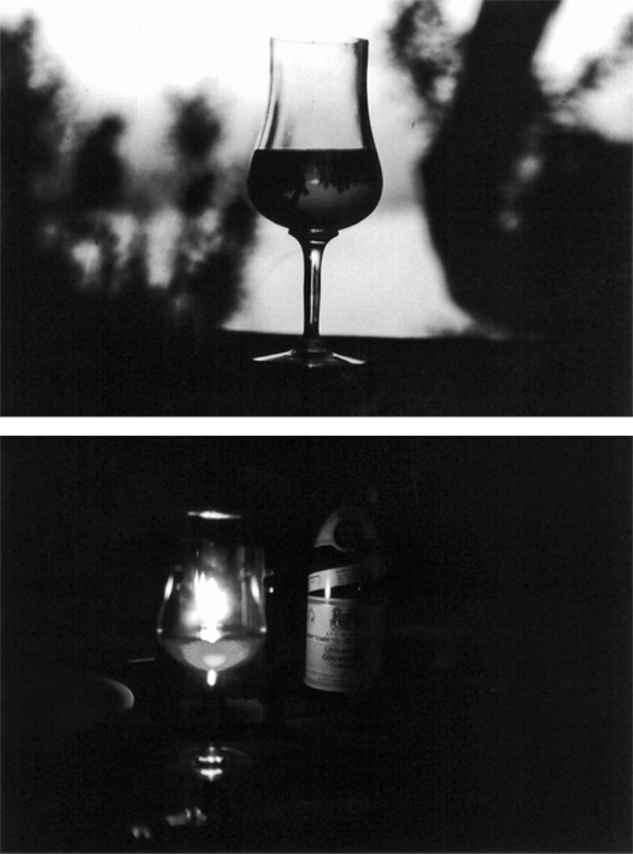
Figure 10-01:
In this picture, a glass is filled with a liquid; however, we do not know what kind of liquid this might be. The bottom picture shows the same glass with a red wine bottle next to it. Although the quality of this image is worse than the top one, we can deduce that the glass contains red wine.
Definition of normal anatomy and pathological changes should be easy and exact. This means that in addition to excellent spatial resolution, high contrast is a prerequisite for a good imaging method.
Magnetic resonance imaging has drawn the attention of many researchers, fascinated by the manifold possibilities of influencing contrast.
In the early years of MR imaging it was believed that image contrast of such quality could be obtained that problems in lesion delineation and even lesion-typing would not occur any more.
The early enthusiasm was rapidly replaced by disillusionment and partial disappointment. It is still not clear whether the method itself is incapable of uncovering all the states and diseases it was intended for or whether poor understanding of the theoretical background of MR imaging led to misguided applications.
Today, many of the early mistakes and misunderstandings can be explained. However, there is enough space for new mistakes. ‘Urban myths’ without sound scientific background appeared and are spreading.
This chapter provides an overview of the main factors and parameters influencing the magnetic resonance image. We will introduce, one by one, the main pulse sequence parameters and see how they influence image contrast.
Contrast in conventional radiographs and CT images is essentially based on small density differences. It can only be changed by adding contrast agents such as barium and iodinated substances that influence electron density within a certain organ.
MR imaging possesses many more contrast-influencing factors and parameters than other imaging methods. One can compare x-ray imaging with radio broadcasting and MR imaging with color television: the former relies on one factor, sound, the latter on sound and moving color pictures. This makes the contrast behavior of MR imaging more complex than that of any other medical imaging modality.
The numerous factors influencing contrast can be divided into two groups: intrinsic and extrinsic parameters. Table 10-01 gives an overview of the most important of these parameters.
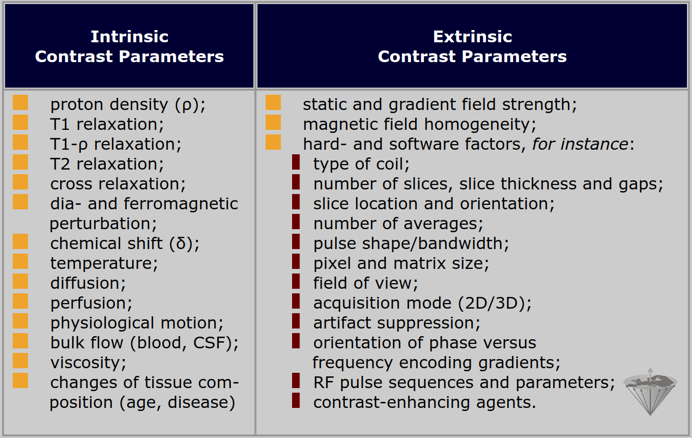
Table 10-01:
Principle intrinsic and extrinsic contrast parameters in magnetic resonance imaging. Please note that this list is long, but not exhaustive.
Many of the extrinsic factors can influence the intrinsic factors. For the clinical application of MR imaging, it is necessary to be aware of all of their interactions if one is to react rapidly and efficiently to a given diagnostic question.
The relative abundance of factors creates a plethora of data, which can impede rather than facilitate the diagnosis, especially if there is a lack of knowledge on how to exploit the information.
One of the main advantages of MR imaging is the possibility to change contrast by choosing special pulse sequences and pulse-sequence parameters.
By emphasizing one factor or mixing several factors in a specific way, the contrast behavior of a certain morphological region or pathological lesion can be highlighted.
One should always bear in mind that even changing minor factors can cause severe contrast changes.
The comparison of two images of the same patient taken with two different machines, apparently using the same parameters, often reveals different contrast patterns.


|

|
|---|