


![]() n the preceding chapters we have discussed the magnetic resonance phenomenon as such, the relaxation times, and the application of magnetic resonance to chemical analysis. However, the most important medical application of magnetic resonance is imaging (Figure 06-01).
n the preceding chapters we have discussed the magnetic resonance phenomenon as such, the relaxation times, and the application of magnetic resonance to chemical analysis. However, the most important medical application of magnetic resonance is imaging (Figure 06-01).
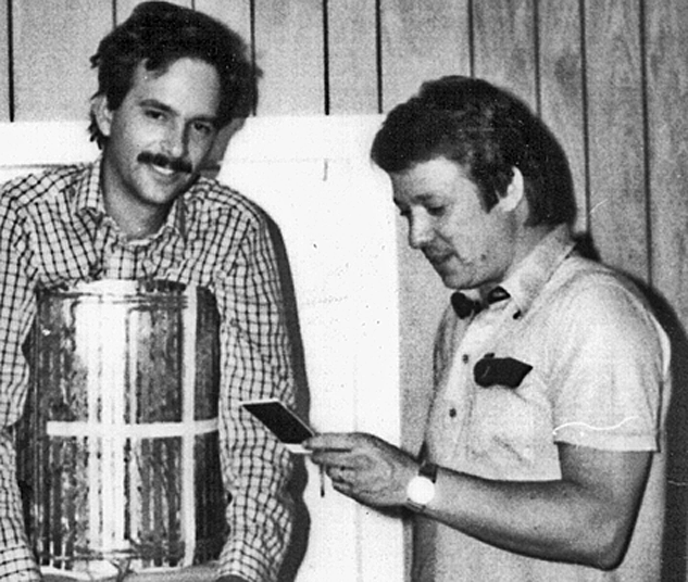
Figure 06-01:
Peter A. Rinck and Robert N. Muller checking a Polaroid print of the first ECG-gated three-dimensional picture of the heart (in 1982). But where do such pictures come from? How are they made?
The manner in which the spatial information is obtained in magnetic resonance imaging is referred to as the reconstruction technique.
Images can be produced point-by-point, line-by-line, in slices, in slices calculated from a whole volume, or as a complete 3D depiction (Figure 06-02).
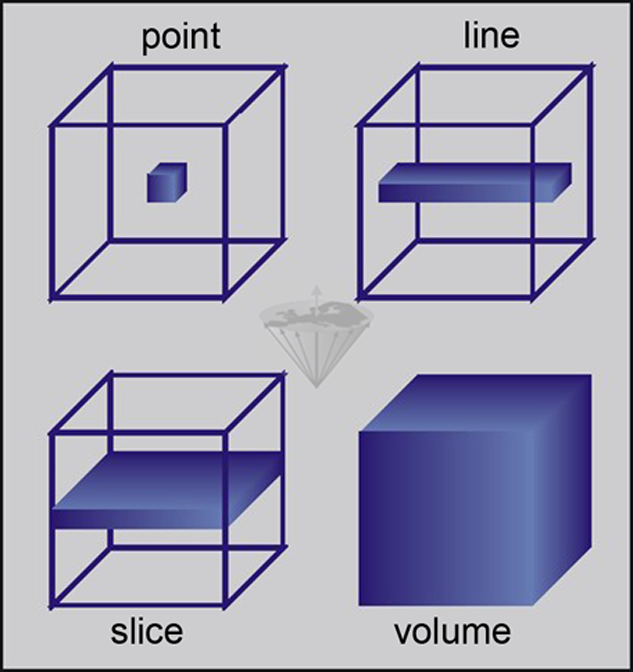
Figure 06-02:
Excitation: point, line, slice, and entire volume.
Nearly all MR imaging techniques currently in use are either planar (slice) or volume techniques. In the former case, the MR experiment is restricted to a slice through the sample and is often referred to as a two-dimensional (2D) experiment since only two spatial dimensions have to be encoded. Volume techniques spatially encode the whole volume; therefore, they are referred to as three-dimensional (3D) techniques.
The formation of an image involves the following procedures:
 localization of the spins of interest;
excitation of selected spins;
spatial encoding of their signal; and
signal detection and reconstruction.


|

|
|---|